Monday, May 17, 2021

Rabies- Lab diagnosis & Prophylaxis

 Lab diagnosis of Rabies:

Human Rabies

-Was of little practical importance till recently- death was considered inevitable.

-Survival was shown possible in rare instances

Diagnosis

A clinical diagnosis of hydrophobia can be made on the basis of history of bite by a rabid animal and characteristic signs and symptoms.

Laboratory diagnosis

Rabies can be confirmed in patients early in the illness by antigen detection using immunofluorescence of skin biopsy, and by virus isolation from saliva and other secretions

1. Demonstration of rabies virus antigens by Immunofluorescence

Specimens: Corneal smears, skin biopsy, (face/neck), saliva -(antemortem) and brain - salivary gland, brain stem, hippocampus, cerebellum - (post mortem)

Direct IF done using anti-rabies serum tagged with fluorescence isothiocyanate.

2. Post-mortem diagnosis by demonstration of Negri bodies in the brain (may be absent in 20% cases)

3. Isolation of Virus by intracerebral inoculation in mice- from brain, CSF, saliva and urine- more chance of isolation early in disease- few days after onset, neutralsiing antibodies appear- inoculated mice examined for signs of illness- brains checked for Negri bodies or by IF

4. Isolation of Virus in tissue culture cell lines (WI 38, BHK 21, CER) – Minimal CPE- virus identified by IF, +ve IF obtained as early as 2-4 days after inoculation

5. High titre antibodies present in CSF in rabies, but not after immunisation- important for diagnosis

6. Detection of rabies virus RNA by RT-PCR- sensitive method, particularly when the sample is small (e.g., saliva) or when large numbers of samples must be tested in an outbreak or epidemiological survey.

Other tests to detect the virus include immunohistochemistry and enzyme-linked immunosorbent assays (ELISAs).

Animal Rabies

Lab diagnosis of rabies in dogs and other biting animals important- to determine risk of infection and to decide post exposure treatment.

In Rabies endemic area, animals captured should be sent for laboratory confirmation of Rabies but without any delay, post exposure treatment of the bitten person should be done.

Domesticated dogs and cats, particularly if previously vaccinated against Rabies should be observed in isolation for upto 10-14 days. If they survive for that time, it is unlikely they were incubating rabies virus at the time of incident. If they succumb or die, anti-rabies treatment of bitten person should be started.

1.     Microscopy/ Histological examination:

This involves the examination of tissue infected with rabies virus rapidly and accurately.

Whole carcass/severed head sent to lab. Brain removed and made into two portions- in 50% glycerol saline (biological test) and 1 in Zenker’s fixative (microscopy). Should include Hippocampus and cerebellum- contain abundant number of Negri bodies.

A definite pathological diagnosis is based on the finding of Negri bodies in the brain or spinal cord. Negri bodies are found in impression preparation or histological sections.

Impression smear- a sample of cells, microorganisms or fluids obtained by pressing against the surface of a specimen, which may be excised tissue or in situ

-Demonstration of Inclusion Bodies-

  • Impression preparation of brain and cornea tissue is often used.

- Brain impression smears stained by Seller’s technique (Basic fuchsin and Methylene Blue in Methanol) – Negri bodies seen as intracytoplasmic, round/oval, purplish pink structures with characteristic basophilic inner granules- vary in size.

·       If impression smears are negative, tissue should be sectioned and stained by Giemsa or Mann’s method.

2. Demonstration of Rabies virus antigen by IF- more sensitive

3. Isolation of Rabies virus- as in human rabies diagnosis

Treatment

There is no specific treatment for rabies, once the clinical signs appear.

Case management includes the following procedure:

(a) The patient should be isolated in a quiet room protected as far as possible from external stimuli such as bright light, noise or cold draughts which may precipitate spasms or convulsions.

(b) Relieve anxiety and pain by liberal use of sedatives.

(d) Ensure hydration and diuresis.

(e) Intensive therapy in the form of respiratory and cardiac support may be given.

 • Patients with rabies are potentially infectious because the virus may be present in the saliva, vomits, tears, urine or other body fluids.

• Nursing personnel attending rabid patients should be warned against possible risk of contamination and should wear face masks, gloves, goggles and aprons to protect themselves. 

• Persons having cuts or open wounds should not look after the patient.

•Where human cases of rabies are encountered frequently, pre-exposure prophylaxis is recommended.

 Prevention

1. Animal Rabies

Rabies can be prevented in domesticated animals by vaccination and by the avoidance of contact with rabid wild animals. Rabies vaccines are available for dogs, cats, cattle, sheep and horses.

Both inactivated and modified live vaccines are effective, but rare cases of post-vaccinal rabies have been reported with the modified live vaccines in dogs and cats.

Preventing animals from roaming will reduce the risk of exposure to rabid wild animals. To protect pet rabbits and rodents, they should be housed indoors, and watched closely if they are allowed outside to exercise.

 2. Human Rabies

a. Post-exposure prophylaxis. b. Pre-exposure prophylaxis.

 Post-Exposure Prophylaxis

The aim of post-exposure prophylaxis is to neutralize the inoculated virus before it can enter the nervous system.

  1. Local treatment of wound: The purpose of local treatment is to remove as much virus as possible from the site of inoculation before it can be absorbed on nerve endings.

• Local treatment of wounds is to be done immediately after exposure; local wound treatment can reduce the chances of developing rabies by up to 80%.

 The local treatment comprises the following measures:

(a) Cleansing: Immediate flushing and washing the wound(s), scratches and the adjoining areas with plenty of soap and water, preferably under a running tap, for at least 15 minutes

• If soap is not available, simple flushing of the wounds with plenty of water should be done as first-aid.

(b) Chemical treatment: Residual virus should be inactivated by irrigation with virucidal agents either alcohol (400-700 ml/litre), tincture or 0.01 % aqueous solution of iodine or Betadine.

(c) Suturing: Bite wounds should not be immediately sutured to prevent additional trauma which may help spread the virus into deeper tissues. If suturing is necessary, it should done 24-48 hours later, applying minimum possible stitches, under the cover of rabies immunoglobulin locally.

(d)Antibiotics and anti-tetanus measure: The application of antibiotics and anti-tetanus procedures when indicated should follow the local treatment recommended above. The use of any local applicant or irritant like turmeric, red chilli, lime etc. should be discouraged and avoided.


  1. Active Immunisation
  • Tissue Culture Vaccines

Human Diploid Cell (HDC) Vaccine

1.     First cell culture vaccine- developed by Koprowski, Wiktor and Plotkin- purified and concentrated preparation of rabies virus (Pitman- Moore strain) grown on human dipoid cells (WI 38 or MRC 5), inactivated with beta propiolactone or tri-n-butyl phosphate- highly antigenic- free from side effects; but has high cost.

2.     Continuous cell culture vaccines grown on the Vero cell line from monkey kidneys

In India, the rabies vaccines available are,

1)     Purified Vero cell rabies vaccine (PVRV)

2)     Chromatographically purified Rabies Vaccine (CPRV)

3)     Human Diploid Cell Vaccine (HDC)

4)     Purified Chick Embryo Cell Vaccine (PCECV) 

5)     Purified Duck Embryo Vaccine (PDEV) 


  • Subunit Vaccine

The Glycoprotein subunit on the virus surface (protective antigen), cloned and recombinant vaccines produced.


Neural vaccines are poor immunogens, contain mostly nucleocapsid antigen- may contain infectious agents and can be encephalitogenic. They are abandoned in most places now, because of the availability of tissue culture vaccines, at affordable price.


Vaccination Schedules

Antirabic vaccine is administered when a person is bitten, scratched or licked by a rabid animal. The animal should be observed for 10 days, if possible. Virus may be present in the saliva for 3-4 days, before the onset of symptoms and the animal usually dies within 5-6 days of developing the disease.

If the animal remains healthy after this period, there is no risk of rabies or vaccination, if already started it may be discontinued.  

WHO guidelines on post-exposure prophylaxis are based on the risk category to which the patient belongs.

 Categories of contact and recommended post-exposure prophylaxis


Categories

Type of contact with suspect rabid animal

Type of exposure

Post–exposure measures

Category I

Touching or feeding animals, licks on intact skin


None

None

Category II

Nibbling of uncovered skin, minor scratches or abrasions without bleeding


Minor

Immediate vaccination (ARV) and local treatment of the wound.

Stop treatment if animal remains healthy throughout an observation period of 10 days or if proven negative for rabies by a reliable laboratory.

Category III

Single or multiple transdermal bites or scratches, licks on broken skin, contamination of mucus membrane with saliva from licks, contact with bats

Severe exposure

Immediate vaccination (ARV) and administration of rabies immunoglobulin, local treatment of the wound.


All three cell culture vaccines available in India (HDC- Human diploid cell vaccine, PCECV - Purified chick embryo cell vaccine, PVRV- Purified vero-cell rabies vaccine) have the same dosage schedule, both for children and adults.

It involves the injection of 0.1 ml of reconstituted vaccine per site and on two sites per visit (one on each deltoid area) on days 0, 3, 7 and 28. Day 0 is the date of the first dose of administration and not the date of exposure/ animal bite.

The vaccine is to be given IM or SC in the deltoid region, or, in the children, on the anterolateral aspect of thigh.


3.     Passive Immunization with antirabies serum

Done by administration of human rabies immunoglobulin (HRIG) pooled from the sera of immunized human donors. Rabies immunoglobulin from the horses (ERIG), was also used, but not generally preferred now due to hypersensitivity reactions. Purified ERIG is much safer, but not completely free from risk. HRIG, though limited in availability and more expensive, is preferred over ERIG, but should be free from HIV and hepatitis viruses.

Administration of Immunoglobulins

All of the rabies immunoglobulin (calculated dose), or as much as anatomically possible should be administered into and around the wound site or sites. The remaining immunoglobulin, if any, after all wound infiltrated, should be administered by deep i/m injection at an injection site distant from the vaccine injection site. Rabies immunoglobulin may be diluted to a volume sufficient (2-3 fold) for all wounds to be effectively and safely infiltrated.

Rabies immunoglobulin for passive immunization is administered only once. Beyond the seventh day after the first dose of ARV, rabies immunoglobulin is not indicated.

Pre exposure prophylaxis:

PEP is recommended for anyone who is at continual, frequent or increased risk of exposure to the rabies virus for example laboratory worker dealing with rabies virus, animal handlers, veterinarians.


Monday, May 10, 2021

Archaea Bacteria - Structure and chemical composition of Archaeal cell wall and cell membranes

The word Archaea is derived from the Greek word Archaios which means ancient.

The Archaeabacteria, are quite diverse, both in morphology and physiology. They may be spherical, rod-shaped, spiral, lobed, cuboidal, triangular, plate-shaped, irregularly shaped, or pleomorphic. Some are single cells, whereas others form filaments or aggregates. They range in diameter from 0.1 to over 15 µm, and some filaments can grow up to 200 µm in length. Multiplication may be by binary fission, budding, fragmentation, or other mechanisms.

The Archaea are diverse physiologically- can be aerobic, facultatively anaerobic, or strictly anaerobic. They include psychrophiles, mesophiles, and hyperthermophiles that can grow above 100°C. Nutritionally they may be chemolithoautotrophs to organotrophs.

Ecology

Archaea are found in areas with either very high or low temperatures or pH, concentrated salts, or completely anoxic. These are generally referred to as “extreme environments.”

Extreme and hypersaline are situations where humans could not survive. Most of the Earth (the oceans) is an “extreme environment” where it is very cold (about 4°C), dark, and under high pressure. Many Archaea are well adapted to these environments, where they can grow to high numbers. Archaea constitute at least 34% of the prokaryotic biomass in some Antarctic coastal waters. In some hypersaline environments, the brine is red with archaeal pigments. Some archaea are symbionts in the digestive tracts of animals. Archaeal gene sequences have been found in soil and temperate and tropical ocean surface waters.

Thus, the Archaea are highly diverse with respect to morphology, reproduction, physiology, and ecology. Although best known for their growth in anoxic, hypersaline, and high-temperature habitats they also inhabit marine arctic, temperate, and tropical waters. Their RNA, ribosomes, elongation factors, RNA polymerases, and other components distinguish Archaea from Bacteria and eukaryotes. Much of archaeal metabolism appears similar to that of other organisms, but the Archaea differ with respect to glucose catabolism, pathways for CO2 fixation, and the ability of some to synthesize methane.

Archaeal Cell Walls

The Archaeal cell wall, like the bacterial cell wall, is a semi-rigid structure which provide protection to the cell from the environment and from the internal cellular pressure. The cell walls of bacteria typically contain peptidoglycan, but it is absent in Archaea. 

Archaeal cell walls stain either Gram positive or Gram negative, depending on the thickness and mass of cell wall. 

The chemistry of Archaeal cell walls is different from that of Eubacteria. Archaea lack the muramic acid and D-amino acids that make up peptidoglycan and thus resist attack by lysozyme and β lactam antibiotics such as penicillin. 

Gram positive Archaea have a variety of complex polymers in their cell wall. Methanobacterium and some other methanogenic archaea have pseudomurein (a peptidoglycan-like polymer that is cross-linked with L-amino acids), N-acetyltalosaminuronic acid instead of N-acetylmuramic acid and β (1- 3) glycosidic bonds instead of β (1-4) glycosidic bonds. Methanosarcina and Halococcus lack pseudomurein and contain complex polysaccharides similar to the chondroitin sulfate of animal connective tissue. Other heteropolysaccharides are also found in Gram positive cellwalls. 

Gram negative Archaea have a layer of protein or glycoprotein (20-40 mm thick) outside their plasma membrane. Some methanogens (Methanolobus), Halobacterium, extreme thermophiles (Sulfolobus, Thermoproteus, Pyrodictium) have glycoproteins in their walls. Other methanogens (Methanococcus, Methanomicrobium, Methanogenium) and extreme thermophile Delsuphurococcus have protein walls.

Structure, function and chemical composition of archaeal cell membranes

One of the most distinctive archaeal features is their membrane lipids. 

Archaeal membrane lipids differ from those of other organisms in having glycerol connected to branched chain hydrocarbons by ether links. 

Bacterial and eukaryotic lipids have glycerol connected to fatty acids by ester bonds.


Sometimes, two glycerol groups are linked to form long tetraethers. Usually, the diether hydrocarbon chains are 20 carbons in length, and the tetraether chains are 40 carbons. Cells adjust the  length of the tetraethers by forming pentacyclic ringsSuch pentacyclic rings are used by thermophilic archaea to help maintain the delicate balance of the membrane at high temperatures. Biphytanyl chains contain 1 to 4 cyclopentyl rings. 

        Phosphate-, sulfur- and sugar-containing groups can be attached to the third carbons of the diethers and tetraethers, making them polar lipids -phospholipids, sulfolipids, and glycolipids.  These predominate in the membrane, making up 70 to 93% of the membrane lipids.  The remaining lipids (7-30%) are nonpolar and are usually derivatives of squalene.


These lipids are combined in different ways to yield membranes of various rigidity and thickness. A regular bilayer membrane is formed when C20 diethers are used. A much more rigid monolayer       membrane is formed when the membrane is constructed of C40 tetraethers. Archaeal membranes may contain a mix of diethers, tetraethers and other lipids. 

The membranes of extreme thermophiles such as Thermoplasma and Sulfolobus contain  tetraether monolayers which provide stability. Archaea that live in moderately hot environments have a mixed membrane containing some regions with monolayers and some with bilayers.


Monday, May 3, 2021

Bacterial Endospore

One of the most common  mechanisms for bacteria against environmental stress is forming endospores. Bacterial spores are the most dormant form of bacteria. They exhibit minimum metabolism, respiration and enzyme production. 

Typically, Gram-positive bacteria are best known for producing intracellular spores called endospores. Endospores are highly refractile (scatter light) and thick-walled structures formed inside the bacterial cells. It is most common for Bacillus species as well as Clostridium species to create endospores. 

Bacterial spores help them to overcome adverse environmental conditions, whereas fungal spores are reproductive in nature.

Endospore is named so, since it is seen within vegetative bacterial cells. It is a dormant and highly resistant structure which helps to preserve the cell's genetic material in times of extreme stress.

Spore forming bacteria - Bacillus and Clostridium (rods), Sporosarcina (cocci).

Spores are,

        resistant (heat, ultraviolet radiation, gamma radiation, chemical disinfectants, and desiccation) & dormant structure

        Viable for around many thousands of years

         Several species of endospore-forming bacteria are dangerous pathogens eg., Bacillus anthracis (Anthrax) and Clostridium tetani (Tetanus)

v Practical importance - food, industrial, and medical microbiology (sterilization problems)

        survive boiling for an hour or more; autoclaves must be used for killing them, if at all

v Theoretical Interest - spore formation -research on the construction of complex biological structures

v In the environment, endospores aid in survival when moisture or nutrients are scarce

Visibility

Examined with light and electron microscopes

v Spores are impermeable to most stains- seen as colorless areas in bacteria treated with simple stains

v special spore stains make them clearly visible- Malachite Green (Primary stain) & Safranin (Counter stain)


v Spore position in the mother cell differs among species; useful in identification

v central, sub terminal – (close to one end ), or terminal

v  some even bulge the sporangium


Spore - Structure


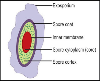
        The spore is surrounded by a thin, delicate covering called the exosporium

        Beneath exosporium, lies the spore coat - fairly thick, made of several protein layers; Impermeable to chemicals - responsible for the spore’s resistance

        Next is the cortex, which is half the spore volume; made of  peptidoglycan (less cross-linked than in vegetative cells)

        The spore cell wall (or core wall) inside the cortex, surrounding the protoplast or core

        The core has the normal cell structures such as ribosomes and a nucleoid, but is metabolically inactive.

Endospore heat resistance contributed by several factors:

Ø  calcium- dipicolinic acid complex

Ø  Stabilization of DNA by acid-soluble proteins

Ø  Dehydrated protoplast

Ø  Spore coat

Ø  DNA repair and others. 

 Ø  Dipicolinic acid complexed with calcium ions - located in the core; contributes to 15% of the spore’s dry weight

       Dipicolinic acid directly involved in spore heat resistance - ( though, heat-resistant mutants lacking dipicolinic acid now have been isolated)

Ø  Calcium aids in resistance to wet and dry heat, oxidizing agents,

        Calcium-dipicolinate stabilizes spore nucleic acids

Ø  Specialized small, acid-soluble DNA-binding proteins in the endospore; stabilize spore DNA and protect it from heat, radiation, desiccation, and chemicals.

Ø  Dehydration of the protoplast - important in heat resistance. The cortex osmotically removes water from the protoplast-protection from both heat and radiation damage

Ø  Spore coat - protect against enzymes and chemicals such as hydrogen peroxide

Ø  DNA repair enzymes – present in spores - which perform repair during germination


Sporogenesis

Ø  Spore formation, sporogenesis or sporulation; when growth ceases due to lack of nutrients.

Ø  Requires only about 10 hours in Bacillus megaterium.

Ø  It is a complex process and may be divided into seven stages


        DNA replicates and aligns along the axis of the cell - axial filament of nuclear material forms (stage I)

        Inward folding of the cell membrane to enclose part of the DNA to produce the forespore septum (stage II)

       The membrane continues to grow and engulfs the immature spore in a second membrane (stage III)

        Cortex laid down in the space between the two membranes, and both calcium and dipicolinic acid are accumulated (stage IV)

        Protein coats then are formed around the cortex (stage V)

        Maturation of the spore occurs (stage VI).

        Lytic enzymes destroy the sporangium releasing the spore (stage VII).


Germination of Spore

Transformation of dormant spores into active vegetative cells - as complex as sporogenesis.  Reactivation of the endospore occurs when conditions are more favourable and involves activation, germination, and outgrowth.

Even if an endospore is located in plentiful nutrients, it may fail to germinate unless activation has taken place. This may be triggered by heating the endospore.

        Three stages: (1) activation, (2) germination, and (3) outgrowth.

        Often an endospore will not germinate successfully, even in a nutrient-rich medium- it has to be activated

        Activation is a reversible process that prepares spores for germination - usually results from treatments like heating

        Germination - the breaking of the spore’s dormant state-      characterized by spore swelling, rupture or absorption of the spore coat, loss of resistance to heat and other stresses, loss of refractility, release of spore components, and increase in metabolic activity;  triggered by many normal metabolites or nutrients (e.g., amino acids and sugars)

        Germination is followed by the third stage, outgrowth-  The spore protoplast makes new components-  emerges from the remains of the spore coat


Thus, germination involves the dormant endospore starting metabolic activity and thus breaking hibernation. It is commonly characterized by rupture or absorption of the spore coat, swelling of the endospore, an increase in metabolic activity, and loss of resistance to environmental stress.

Outgrowth follows germination and involves the core of the endospore manufacturing new chemical components and exiting the old spore coat to develop into a fully functional vegetative bacterial cell, which can divide to produce more cells.

Endospores can stay dormant for a very long time. For instance, endospores were found in the tombs of the Egyptian pharaohs. When placed in appropriate medium, under appropriate conditions, they were able to be reactivated.

 In 1995, Raul Cano of California Polytechnic State University found bacterial spores in the gut of a fossilized bee trapped in amber from a tree in the Dominican Republic. The bee fossilized in amber was dated to being about 25 million years old. The spores germinated when the amber was cracked open and the material from the gut of the bee was extracted and placed in nutrient medium.


Effect of aeration on growth of bacteria

  Aim To study the effect of aeration on growth of bacteria Principle Microorganisms are classified into categories such as aerobic, f...