Staphylococci are gram positive cocci that appear in grape like clusters. They are ubiquitous, and are the most common cause of localized suppurative (pus containing) lesions in humans. They are aerobes/facultative anaerobes, catalase positive bacteria, comprising about 40 species and subspecies.
They develop resistance to penicillin and other antibiotics easily and hence are important human pathogens especially in the hospital environment (nosocomial pathogens).
Staphylococci were first observed in human pus in 1871 by Von Recklinghausen.
In 1880, Louis Pasteur first cultured it in liquid media and inoculated them in rabbits and observed abscesses.
It was named “Staphylococcus” (Staphyle (G) = bunch of grapes; kokkos=berry) by Sir Alexander Ogston, a Scottish surgeon in 1880. He established conclusively the role of Staphylococci in abscesses and suppurative lesions. He also noticed the presence of nonvirulent Staphylococci on skin surfaces.
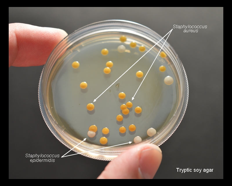
In 1884, Rosenbach identified and named two strains based on their growth on solid media. - Moist virulent Staphylococci from the lesions were found to produce golden yellow colonies hence named Staphylococcus aureus (golden yellow colonies)
- the strains from normal skin, produced white colonies on solid media, hence named Staphylococcus albus,
-In 1885, Passet identified a third variety, S. citreus producing lemon yellow colonies.
Classification is:
Genus - Staphylococcus
Family - Micrococcaceae
Species - Staphylococcus aureus, S. citreus, S. albus etc.,
Staphylococci are classified into two groups based on pigment production and some other biochemical characters, especially presence of Coagulase enzyme and fermentation of mannitol, two characters which was found to have some association with its virulence.
Staphylococcus aureus (Staphylococcus pyogenes, earlier) and similar strains are pathogenic, coagulase positive and ferment mannitol. Staphylococcus epidermidis (former Staphylococcus albus), and similar strains are usually non-pathogenic, coagulase negative and do not ferment mannitol.
However, three species among them, S. epidermidis, S. haemolyticus, S. saprophyticus, are found to cause human diseases. Some other coagulase negative species like S. hominis and S. capitus are part of commensal flora of human skin. Other species are parasitic on animals.
Briefly,
Genus Staphylococci Divided into 2 sub-groups especially based on the presence of Enzyme coagulase
1. Genus Staphylococcus A- Coagulase positive Staphylococci - Staphylococcus aureus, Staphylococcus intermedius, Staphylococcus hyicus
2. Genus Staphylococcus B- Coagulase Negative Staphylococci (CONS) - Staphylococcus epidermidis (Staphylococcus albus), Staphylococcus citreus, Staphylococcus saprophyticus, Staphylococcus hominis, Staphylococcus capitus, Staphylococcus hemolyticus.
Staphylococcus genus includes at least 40 species
The three main species of clinical importance are:
• S. aureus: pathogenic and commensally found in nose (nares).
• S. epidermidis: is a commensal of the skin; can cause severe infections in immune-suppressed patients.
• S. saprophyticus: is part of the normal vaginal flora, implicated in genitourinary tract infections.
Staphylococcus aureus
Morphology
Spherical, Gram positive cocci in irregular grape like clusters. Arranged characteristically, in grape like clusters due to the cell division occurring in three planes and the daughter cells remaining attached to one another. Also found singly, in pairs or short chains of three or four cells, especially when observed from liquid culture.

They are non-motile, non-sporing, usually non-capsulated (Few strains possess microscopically visible capsule, especially in young cultures). They stain readily with aniline dye & are always Gram Positive. Peptidoglycan layer is the major structural component of the cell wall. Teichoic acid is present. They occur as L forms in the presence of penicillin and certain cell wall lysing chemicals.
Cultural
Characters
Aerobes and facultative
anaerobes, Grow readily on ordinary media with an optimum growth temperature of
37°C and optimum pH 7.4 - 7.6.
On Nutrient agar, after 24 h incubation, they form large (2-4 mm diameter), circular, convex, golden yellow opaque, easily emulsifiable colonies with smooth shiny/glistening surface. The pigment does not diffuse into the medium and maximum pigment production occurs at 22 °C, in aerobic conditions. Pigment production can be enhanced by adding 1% glycerol monoacetate or milk in the medium.

On Blood agar, similar golden
yellow colonies, surrounded by a clear zone of hemolysis (β-hemolysis /clear
zone around the colonies) when incubated under 20-25% CO2. Hemolysis is pronounced in sheep or rabbit
blood and weak on horse blood agar.

(S. epidermidis -White-creamy colonies -no hemolysis of red blood cells.
S. Saprophyticus - white-yellow colony -no hemolysis of red blood cells)
On MacConkey’s agar, they produce smaller colonies (0.1-0.5 mm) than those on nutrient agar and are pink coloured due to lactose fermentation.

On Mannitol salt agar, S. aureus ferments mannitol and appear as yellow colonies. MSA is a useful selective/differential medium for recovering S. aureus from faecal specimens, when investigating food poisoning.

Growth on other media:
1. Liquid media: uniform turbidity
2. Selective
media for isolating S.
aureus (from faecal specimens etc)
ü Milk salt agar
or broth – contain 8-10% NaCl
ü Mannitol salt agar –
contain Mannitol
ü Baird – Parker
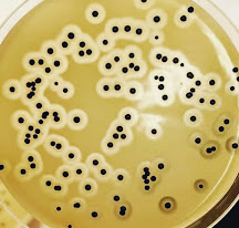
agar containing Polymyxin B ( selective and diagnostic medium for coagulase-positive staphylococci in foods-a good differentiation medium for coagulase positive strains-egg yolk emulsion as a differentiation agent-of a clear zone of lipolysis due to the lecithinase of Staphylococci that breakdown, the egg yolk - coagulase positive Staphylococci can reducing
tellurite and produce black colonies, whereas other Staphylococci
cannot always)
ü Ludlam’s media- Lithium chloride, Tellurite with Polymyxin B (Lithium chloride and potassium tellurite inhibit contaminating microflora except Staphylococcus aureus)
ü For Primary isolation, Sheep Blood Agar Plate (S-BAP) recommended – human blood avoided since it contains antibodies/inhibitors
- Biochemical properties
- • Ferment glucose, lactose, maltose, sucrose and mannitol, with production of acid but no gas
- • Mannitol fermentation by S. aureus, and no other species- has diagnostic significance
- • Catalase positive
- • Urease positive (Hydrolyse Urea)
- • Reduces nitrate to nitrite
- • Indole negative
- • MR positive
- • VP positive
- • Hydrolyse gelatin
- •Phosphatase positive (S, epidermidis is negative/weakly positive)
- • DNA-ase test positive - Produce thermostable nucleases (demonstrated by the ability of boiled cultures to degrade DNA in an agar diffusion test)
- • Coagulase test positive
- • Oxidase negative
- •Clear hemolysis on Blood agar
- • Golden yellow pigment
- •Produce black colonies on a medium containing potassium tellurite,by reducing tellurite.


No comments:
Post a Comment